Gastrointestinal Foreign Bodies: Background, Pathophysiology, Epidemiology
Gastrointestinal Foreign Bodies: Background, Pathophysiology, Epidemiology
Patients with foreign bodies in the gastrointestinal (GI) tract commonly present to the ED. Foreign bodies in the upper GI tract are usually swallowed, purposefully or accidentally. The presentation is usually straightforward but on occasion can be extremely subtle. A foreign body in the GI tract is shown in the radiograph below.
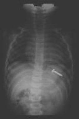 A screw in the stomach; peristaltic action will carry the screw through the GI tract with the blunt end (head) leading and the sharp end trailing.
Most of the literature covering GI foreign bodies is anecdotal, with the exception of some recent studies on esophageal foreign body removal techniques.
Foreign bodies may involve the entire upper GI tract. The oropharynx is well innervated, and patients can typically localize oropharyngeal foreign bodies. Scratches or abrasions to the mucosal surface of the oropharynx can create a foreign body sensation. Chronic foreign bodies or perforations can cause infections in surrounding soft tissues of the throat and neck.
The esophagus is a tubular structure approximately 20-25 cm in length. Patients can usually localize foreign bodies in the upper esophagus but localize them poorly in the lower two thirds of the structure. The esophagus has 3 areas of narrowing where foreign bodies are most likely to become entrapped: the upper esophageal sphincter (UES), which consists of the cricopharyngeus muscle; the crossover of the aorta; and the lower esophageal sphincter (LES). Structural abnormalities of the esophagus, including strictures, webs, diverticula, and malignancies, increase the risk of foreign body entrapment, as do motor disturbances such as scleroderma, diffuse esophageal spasm, or achalasia.
After reaching the stomach, a foreign body has greater than a 90% chance of passage. Coins reaching the stomach are very likely to pass into the small bowel. Objects larger than 2 cm in diameter are less likely to pass the pylorus, and objects longer than 6 cm may become entrapped at either the pylorus or the duodenal sweep. Objects reaching the small bowel occasionally are impeded by the ileocecal valve. Rarely, a foreign body may become entrapped in a Meckel diverticulum.
Swallowed magnets from toys and household items have become a serious health hazard in children. Buckey-ball magnets are small round magnets in the shape of ball-bearings that are especially strong and are used to make toys of various shapes. If these small magnets are ingested, especially at various times, they can adhere across layers of bowel and lead to pressure necrosis, fistula, volvulus, perforation, infection, or obstruction.
United States
The incidence of foreign body ingestions in children and adults is unknown. Data are largely anecdotal.
A recent study suggested approximately 1671 ingested magnet injuries annually. This is expected to decrease since sales of these small toy magnets have been banned by the Consumer Protection Agency because of safety concerns.
An estimated 1500 deaths occur annually from foreign bodies in the upper GI tract.
Complications
Complications of GI foreign bodies include the following:
No differences in race or nationality have been noted.
In children with swallowed foreign bodies, the incidence in males and females is equal.In adults, the incidence of accidentally swallowed foreign bodies is slightly higher in men than in women, and the incidence of intentionally swallowed foreign bodies is much higher in men than in women.
Patients with foreign bodies in the upper GI tract usually fall into 1 of 3 categories: (1) children, (2) psychiatric patients and prisoners, and (3) edentulous patients.
Children account for 75-85% of patients with foreign bodies in the upper GI tract, with a preponderance at age 18-48 months.
The objects involved also differ by group. Children typically ingest objects they pick up and place in their mouths, such as coins, buttons, marbles, crayons, and similar items.In contrast, adults are more prone to ingest food boluses, chicken or fish bones, fruit pits, dentures, or toothpicks.Prisoners and psychiatric patients may present with bizarre objects, as well as multiple objects.
The site of entrapment of esophageal foreign bodies also differs with age groups, with about 75% of children having entrapment at the upper esophageal sphincter (UES) and about 70% of adults having entrapment at the lower esophageal sphincter (LES).
Clinical Presentation
David W Munter, MD, MBA Associate Clinical Professor of Emergency Medicine, Eastern Virginia Medical School; Associate Professor of Emergency Medicine, Edward Via Virginia College of Osteopathic Medicine; Partner, Emergency Physicians of Tidewater, PLC; President of the DESA Consulting Group
David W Munter, MD, MBA is a member of the following medical societies: American College of Emergency Physicians, Medical Society of Virginia, Norfolk Academy of Medicine, American Association for Physician Leadership
Specialty Editor Board
Francisco Talavera, PharmD, PhD Adjunct Assistant Professor, University of Nebraska Medical Center College of Pharmacy; Editor-in-Chief, Medscape Drug Reference
Disclosure: Received salary from Medscape for employment. for: Medscape.
Chief Editor
Steven C Dronen, MD, FAAEM Chair, Department of Emergency Medicine, LeConte Medical Center
Steven C Dronen, MD, FAAEM is a member of the following medical societies: American Academy of Emergency Medicine, Society for Academic Emergency Medicine
Jerry R Balentine, DO, FACEP, FACOEP Vice President, Medical Affairs and Global Health, New York Institute of Technology; Professor of Emergency Medicine, New York Institute of Technology College of Osteopathic Medicine
Jerry R Balentine, DO, FACEP, FACOEP is a member of the following medical societies: American College of Emergency Physicians, New York Academy of Medicine, American College of Osteopathic Emergency Physicians, American Association for Physician Leadership, American Osteopathic Association
Acknowledgements
Eugene Hardin, MD, FAAEM, FACEP Former Chair and Associate Professor, Department of Emergency Medicine, Charles Drew University of Medicine and Science; Former Chair, Department of Emergency Medicine, Martin Luther King Jr/Drew Medical Center
References
Coin (quarter) lodged at the level of the cricopharyngeus muscle.
Coin lodged at the level of the aortic crossover.
Coin lodged at the lower esophageal sphincter.
A screw in the stomach; peristaltic action will carry the screw through the GI tract with the blunt end (head) leading and the sharp end trailing.
Background
Patients with foreign bodies in the gastrointestinal (GI) tract commonly present to the ED. Foreign bodies in the upper GI tract are usually swallowed, purposefully or accidentally. The presentation is usually straightforward but on occasion can be extremely subtle. A foreign body in the GI tract is shown in the radiograph below.
A screw in the stomach; peristaltic action will carry the screw through the GI tract with the blunt end (head) leading and the sharp end trailing. Most of the literature covering GI foreign bodies is anecdotal, with the exception of some recent studies on esophageal foreign body removal techniques.
Pathophysiology
Foreign bodies may involve the entire upper GI tract. The oropharynx is well innervated, and patients can typically localize oropharyngeal foreign bodies. Scratches or abrasions to the mucosal surface of the oropharynx can create a foreign body sensation. Chronic foreign bodies or perforations can cause infections in surrounding soft tissues of the throat and neck.
The esophagus is a tubular structure approximately 20-25 cm in length. Patients can usually localize foreign bodies in the upper esophagus but localize them poorly in the lower two thirds of the structure. The esophagus has 3 areas of narrowing where foreign bodies are most likely to become entrapped: the upper esophageal sphincter (UES), which consists of the cricopharyngeus muscle; the crossover of the aorta; and the lower esophageal sphincter (LES). Structural abnormalities of the esophagus, including strictures, webs, diverticula, and malignancies, increase the risk of foreign body entrapment, as do motor disturbances such as scleroderma, diffuse esophageal spasm, or achalasia.
After reaching the stomach, a foreign body has greater than a 90% chance of passage. Coins reaching the stomach are very likely to pass into the small bowel. Objects larger than 2 cm in diameter are less likely to pass the pylorus, and objects longer than 6 cm may become entrapped at either the pylorus or the duodenal sweep. Objects reaching the small bowel occasionally are impeded by the ileocecal valve. Rarely, a foreign body may become entrapped in a Meckel diverticulum.
Swallowed magnets from toys and household items have become a serious health hazard in children. Buckey-ball magnets are small round magnets in the shape of ball-bearings that are especially strong and are used to make toys of various shapes. If these small magnets are ingested, especially at various times, they can adhere across layers of bowel and lead to pressure necrosis, fistula, volvulus, perforation, infection, or obstruction.
Epidemiology
Frequency
United States
The incidence of foreign body ingestions in children and adults is unknown. Data are largely anecdotal.
A recent study suggested approximately 1671 ingested magnet injuries annually. This is expected to decrease since sales of these small toy magnets have been banned by the Consumer Protection Agency because of safety concerns.
Mortality/Morbidity
An estimated 1500 deaths occur annually from foreign bodies in the upper GI tract.
Complications
Complications of GI foreign bodies include the following:
- Potential complications of oropharyngeal foreign bodies include esophageal or pharyngeal abrasions, lacerations, and punctures, with associated abscesses (eg, retropharyngeal abscess), perforations, and soft-tissue infections.
- Esophageal foreign bodies can also cause mucosal scratches or abrasions, punctures, and perforations, with resultant injuries or infections to surrounding structures, including abscesses, pneumomediastinum or mediastinitis ; pericarditis/tamponade, pneumothorax, pneumomediastinum, tracheoesophageal fistula, or even vascular injuries to the aorta (aortoesophageal fistulas) ; or pulmonary vasculature. Additionally, button batteries can rapidly create esophageal necrosis. Esophageal strictures may also occur.
- Complications from foreign bodies in the stomach and small intestine typically involve perforation and associated infection, including peritonitis and sepsis. Small-bowel obstruction may also occur
- Swallowed toy magnets that adhere across layers of bowel can cause pressure necrosis, fistula, volvulus, perforation, infection, or obstruction.
Race
No differences in race or nationality have been noted.
Sex
In children with swallowed foreign bodies, the incidence in males and females is equal.In adults, the incidence of accidentally swallowed foreign bodies is slightly higher in men than in women, and the incidence of intentionally swallowed foreign bodies is much higher in men than in women.
Age
Patients with foreign bodies in the upper GI tract usually fall into 1 of 3 categories: (1) children, (2) psychiatric patients and prisoners, and (3) edentulous patients.
Children account for 75-85% of patients with foreign bodies in the upper GI tract, with a preponderance at age 18-48 months.
The objects involved also differ by group. Children typically ingest objects they pick up and place in their mouths, such as coins, buttons, marbles, crayons, and similar items.In contrast, adults are more prone to ingest food boluses, chicken or fish bones, fruit pits, dentures, or toothpicks.Prisoners and psychiatric patients may present with bizarre objects, as well as multiple objects.
The site of entrapment of esophageal foreign bodies also differs with age groups, with about 75% of children having entrapment at the upper esophageal sphincter (UES) and about 70% of adults having entrapment at the lower esophageal sphincter (LES).
Clinical Presentation
David W Munter, MD, MBA Associate Clinical Professor of Emergency Medicine, Eastern Virginia Medical School; Associate Professor of Emergency Medicine, Edward Via Virginia College of Osteopathic Medicine; Partner, Emergency Physicians of Tidewater, PLC; President of the DESA Consulting Group
David W Munter, MD, MBA is a member of the following medical societies: American College of Emergency Physicians, Medical Society of Virginia, Norfolk Academy of Medicine, American Association for Physician Leadership
Specialty Editor Board
Francisco Talavera, PharmD, PhD Adjunct Assistant Professor, University of Nebraska Medical Center College of Pharmacy; Editor-in-Chief, Medscape Drug Reference
Disclosure: Received salary from Medscape for employment. for: Medscape.
Chief Editor
Steven C Dronen, MD, FAAEM Chair, Department of Emergency Medicine, LeConte Medical Center
Steven C Dronen, MD, FAAEM is a member of the following medical societies: American Academy of Emergency Medicine, Society for Academic Emergency Medicine
Jerry R Balentine, DO, FACEP, FACOEP Vice President, Medical Affairs and Global Health, New York Institute of Technology; Professor of Emergency Medicine, New York Institute of Technology College of Osteopathic Medicine
Jerry R Balentine, DO, FACEP, FACOEP is a member of the following medical societies: American College of Emergency Physicians, New York Academy of Medicine, American College of Osteopathic Emergency Physicians, American Association for Physician Leadership, American Osteopathic Association
Acknowledgements
Eugene Hardin, MD, FAAEM, FACEP Former Chair and Associate Professor, Department of Emergency Medicine, Charles Drew University of Medicine and Science; Former Chair, Department of Emergency Medicine, Martin Luther King Jr/Drew Medical Center
References
- Silverman JA, Brown JC, Willis MM, Ebel BE. Increase in pediatric magnet-related foreign bodies requiring emergency care. Ann Emerg Med. 2013 Dec. 62(6):604-608.e1. [Medline].
- Stack LB, Munter DW. Foreign bodies in the gastrointestinal tract. Emerg Med Clin North Am. 1996 Aug. 14(3):493-521. [Medline].
- Ghimire A, Bhattarai M, Kumar M, Wakode PT. Descending necrotizing mediastinitis: a fatal complication of neglected esophageal foreign body. Kathmandu Univ Med J (KUMJ). 2007 Jan-Mar. 5(1):98-101. [Medline].
- Macchi V, Porzionato A, Bardini R, Parenti A, De Caro R. Rupture of ascending aorta secondary to esophageal perforation by fish bone. J Forensic Sci. 2008 Sep. 53(5):1181-4. [Medline].
- Kunishige H, Myojin K, Ishibashi Y, Ishii K, Kawasaki M, Oka J. Perforation of the esophagus by a fish bone leading to an infected pseudoaneurysm of the thoracic aorta. Gen Thorac Cardiovasc Surg. 2008 Aug. 56(8):427-9. [Medline].
- Balci AE, Eren S, Eren MN. Esophageal foreign bodies under cricopharyngeal level in children: an analysis of 1116 cases. Interact Cardiovasc Thorac Surg. 2004 Mar. 3(1):14-8. [Medline].
- Nadir A, Sahin E, Nadir I, Karadayi S, Kaptanoglu M. Esophageal foreign bodies: 177 cases. Dis Esophagus. 2011 Jan. 24(1):6-9. [Medline].
- Hurtado CW, Furuta GT, Kramer RE. Etiology of esophageal food impactions in children. J Pediatr Gastroenterol Nutr. 2011 Jan. 52(1):43-6. [Medline].
- Conway WC, Sugawa C, Ono H, Lucas CE. Upper GI foreign body: an adult urban emergency hospital experience. Surg Endosc. 2007 Mar. 21(3):455-60. Epub 2006 Nov 28. [Medline].
- Chinski A, Foltran F, Gregori D, Ballali S, Passali D, Bellussi L. Foreign Bodies in the Oesophagus: The Experience of the Buenos Aires Paediatric ORL Clinic. Int J Pediatr. 2010. 2010:[Medline]. [Full Text].
- Baral BK, Joshi RR, Bhattarai BK, Sewal RB. Removal of coin from upper esophageal tract in children with Magill's forceps under propofol sedation. Nepal Med Coll J. 2010 Mar. 12(1):38-41. [Medline].
- Little DC, Shah SR, St Peter SD, Calkins CM, Morrow SE, Murphy JP, et al. Esophageal foreign bodies in the pediatric population: our first 500 cases. J Pediatr Surg. 2006 May. 41(5):914-8. [Medline].
- Louie JP, Alpern ER, Windreich RM. Witnessed and unwitnessed esophageal foreign bodies in children. Pediatr Emerg Care. 2005 Sept. 21(9):582-5. [Medline].
- Kim N, Atkinson N, Manicone P. Esophageal foreign body: a case of a neonate with stridor. Pediatr Emerg Care. 2008 Dec. 24(12):849-51. [Medline].
- Miller RS, Willging JP, Rutter MJ, Rookkapan K. Chronic esophageal foreign bodies in pediatric patients: a retrospective review. Int J Pediatr Otorhinolaryngol. 2004 Mar. 68(3):265-72. [Medline].
- Lee SC, Ebert CS Jr, Fordham L, Rose AS. Plain films in the evaluation of batteries as esophageal foreign bodies. Int J Pediatr Otorhinolaryngol. 2008 Oct. 72(10):1487-91. [Medline].
- Hergan K, Kofler K, Oser W. Drug smuggling by body packing: what radiologists should know about it. Eur Radiol. 2004 Apr. 14(4):736-42. Epub 2003 Oct 18. [Medline].
- Palme CE, Lowinger D, Petersen AJ. Fish bones at the cricopharyngeus: a comparison of plain-film radiology and computed tomography. Laryngoscope. 1999 Dec. 109(12):1955-8. [Medline].
- Eliashar R, Dano I, Dangoor E, Braverman I, Sichel JY. Computed tomography diagnosis of esophageal bone impaction: a prospective study. Ann Otol Rhinol Laryngol. 1999 Jul. 108(7 Pt 1):708-10. [Medline].
- Bassett KE, Schunk JE, Logan L. Localizing ingested coins with a metal detector. Am J Emerg Med. 1999 Jul. 17(4):338-41. [Medline].
- Chung CH, Fung WT. Detection of gastric drug packet by ultrasound scanning. Eur J Emerg Med. 2006 Oct. 13(5):302-3. [Medline].
- Lin HH, Lee SC, Chu HC, Chang WK, Chao YC, Hsieh TY. Emergency endoscopic management of dietary foreign bodies in the esophagus. Am J Emerg Med. 2007 Jul. 25(6):662-5. [Medline].
- Lin CH, Chen AC, Tsai JD, Wei SH, Hsueh KC, Lin WC. Endoscopic removal of foreign bodies in children. Kaohsiung J Med Sci. 2007 Sep. 23(9):447-52. [Medline].
- Pokharel R, Adhikari P, Bhusal CL, Guragain RP. Oesophageal foreign bodies in children. JNMA J Nepal Med Assoc. 2008 Oct-Dec. 47(172):186-8. [Medline].
- Lue AJ, Fang WD, Manolidis S. Use of plain radiography and computed tomography to identify fish bone foreign bodies. Otolaryngol Head Neck Surg. 2000 Oct. 123(4):435-8. [Medline].
- Litovitz T, Whitaker N, Clark L, White NC, Marsolek M. Emerging battery-ingestion hazard: clinical implications. Pediatrics. 2010 Jun. 125(6):1168-77. [Medline].
- Litovitz T, Whitaker N, Clark L. Preventing battery ingestions: an analysis of 8648 cases. Pediatrics. 2010 Jun. 125(6):1178-83. [Medline].
- Sharpe SJ, Rochette LM, Smith GA. Pediatric Battery-Related Emergency Department Visits in the United States,1999-2009. Pediatrics. 5/14/2012. doi:10.1542:[Full Text].
- Waltzman ML. Management of esophageal coins. Curr Opin Pediatr. 2006 Oct. 18(5):571-4. [Medline].
- Calkins CM, Christians KK, Sell LL. Cost analysis in the management of esophageal coins: endoscopy versus bougienage. J Pediatr Surg. 1999 Mar. 34(3):412-4. [Medline].
- Bonadio WA, Jona JZ, Glicklich M, Cohen R. Esophageal bougienage technique for coin ingestion in children. J Pediatr Surg. 1988 Oct. 23(10):917-8. [Medline].
- Dashan AH, Kevin Donovan G. Bougienage versus endoscopy for esophageal coin removal in children. J Clin Gastroenterol. 2007 May-Jun. 41(5):454-6. [Medline].
- Cetinkursun S, Sayan A, Demirbag S, Surer I, Ozdemir T, Arikan A. Safe removal of upper esophageal coins by using Magill forceps: two centers' experience. Clin Pediatr (Phila). 2006 Jan-Feb. 45(1):71-3. [Medline].
- Bhargava R, Brown L. Esophageal coin removal by emergency physicians: a continuous quality improvement project incorporating rapid sequence intubation. CJEM. 2011 Jan. 13(1):28-33. [Medline].
- Al-Haddad M, Ward EM, Scolapio JS, Ferguson DD, Raimondo M. Glucagon for the relief of esophageal food impaction does it really work?. Dig Dis Sci. 2006 Nov. 51(11):1930-3. [Medline].
- Metha D, Attia M, Cronan K. Glucagon for esophageal coin dislodgement in children: a prospective, double-blind, placebo-controlled study. Acad Emerg Med. 2001 Feb. 8(2):200-3. [Medline].
- Leopard D, Fishpool S, Winter S. The management of oesophageal soft food bolus obstruction: a systematic review. Ann R Coll Surg Engl. 2011 Sep. 93(6):441-4. [Medline].
- Conners GP. A literature-based comparison of three methods of pediatric esophageal coin removal. Pediatr Emerg Care. 1997 Apr. 13(2):154-7. [Medline].
- Arms JL, Mackenberg-Mohn MD, Bowen MV, Chamberlain MC, Skrypek TM, Madhok M, et al. Safety and efficacy of a protocol using bougienage or endoscopy for the management of coins acutely lodged in the esophagus: a large case series. Ann Emerg Med. 2008 Apr. 51(4):367-72. [Medline].
- Conners GP, Chamberlain JM, Ochsenschlager DW. Conservative management of pediatric distal esophageal coins. J Emerg Med. 1996 Nov-Dec. 14(6):723-6. [Medline].
- Soprano JV, Fleisher GR, Mandl KD. The spontaneous passage of esophageal coins in children. Arch Pediatr Adolesc Med. 1999 Oct. 153(10):1073-6. [Medline].
- Conners GP, Chamberlain JM, Ochsenschlager DW. Symptoms and spontaneous passage of esophageal coins. Arch Pediatr Adolesc Med. 1995 Jan. 149(1):36-9. [Medline].
- Weiland ST, Schurr MJ. Conservative treatment of ingested foreign bodies. J Gastrointest Surg. 2002 May-Jun. 6(3):496-500. [Medline].
- Durko A, Czkwianianc E, Bak-Romaniszyn L, Malecka-Panas E. [Accidental ingestion of two magnets--aggresive or prolonged approach?]. Pol Merkur Lekarski. 2007 May. 22(131):416-8. [Medline].
- Baliga SK, Hussain D, Sarfraz SL, Hartung RU. Magnetic attraction: dual complications in a single case. J Coll Physicians Surg Pak. 2008 Jul. 18(7):440-1. [Medline].
- Cortes C, Silva C. [Accidental ingestion of magnets in children. Report of three cases]. Rev Med Chil. 2006 Oct. 134(10):1315-9. [Medline].
- Hou SK, Chern CH, How CK, Wang LM, Huang CI, Lee CH. Press through package mis-swallowing. Int J Clin Pract. 2006 Feb. 60(2):234-7. [Medline].
- Lee KI, Wang CP. Images in emergency medicine. Press-through package mis-swallowing. Ann Emerg Med. 2008 Aug. 52(2):98, 115. [Medline].
- Butterworth JR, Wright K, Boulton RA, Pathmakanthan S, Goh J. Management of swallowed razor blades-retrieve or wait and see?. Gut. 2004 Apr. 53(4):477, 486. [Medline].
Coin (quarter) lodged at the level of the cricopharyngeus muscle.
Coin lodged at the level of the aortic crossover.
Coin lodged at the lower esophageal sphincter.
A screw in the stomach; peristaltic action will carry the screw through the GI tract with the blunt end (head) leading and the sharp end trailing.
Source...